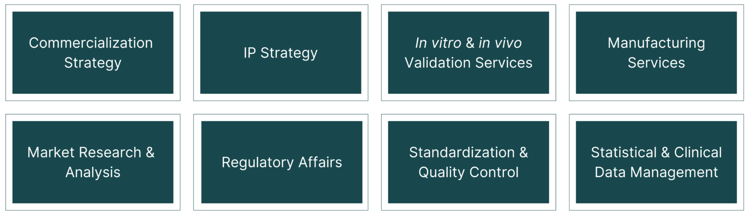
As part of the C-DOCTOR ITP Program, ITP teams can access Core Services and Resources to address their translational research needs in the following areas:
The C-DOCTOR Internal Leadership Council, Operations Committee, NIDCR program staff, and the DOCTRC External Advisory board assess ITP team progress on a quarterly basis to provide feedback, address pitfalls, develop alternative approaches, and implement countermeasure strategies to strengthen the likelihood of securing IND/IDE approval from the FDA to launch clinical trials, as well as future commercialization and clinical adoption.
Consultants and CROs/CMOs in the C-DOCTOR network:
C-DOCTOR brings together uniquely comprehensive resources and infrastructure to support Interdisciplinary Translational Project teams throughout the product development cycle from conception to commercialization. The Director of our Core Resources is Dr. Michael Jamieson. Resource teams include:
Animal models
UC Davis Department of Animal Science
Large and small animal models
USC Department of Animal Resources – James Finlay (jbfinlay@med.usc.edu)
Large and small animals
USC Transgenic/Knockout Rodent Core Facility (Genome Modification Facility) – Yuntao Wang (yuntaow@usc.edu)
Transgenic & knockout mice, CRISPR, knockout rats
UCLA Zebrafish Core – Alvaro Sagasti (zebrafish@ucla.edu)
Transgenesis, large-scale genetic screens
UCLA Mouse Physiology Lab – Kenneth Roos (kroos@mednet.ucla.edu)
Electrocardiography, hemodynamics, surgical procedures, telemetric recording, autonomic testing, exercise & metabolic testing, postmortem morphometry, isolated cell studies, certification testing of new drugs
UCLA Behavioral Testing Core – (uclabtc@gmail.com)
Behavioral testing of mice and rats
UCLA Center for AIDS Research Humanized Mouse Core – Scott Kitchen (skitchen@ucla.edu)
Humanized immunodeficient mice
Biochemistry
Stanford Medicinal Chemistry Knowledge Center – Mark Smith (mxsmith@stanford.edu)
Provides help to biologists and clinicians to incorporate medicinal chemistry into their research endeavors
Stanford Macromolecular Structure Knowledge Center – Daniel Fernandez (danilo@stanford.edu)
Production, purification, and characterization of biological macromolecules
Stanford Metabolomics Knowledge Center – Yuqin Dai (yuqindai@stanford.edu)
Measurement of metabolites
UCLA Biological Chemistry Imaging Facility
Fluorescent scanning equipment and gel documentation systems
UCLA Protein Expression Technology Center – Mark Arbing (marbing@mbi.ucla.edu)
All aspects of protein expression from cloning through expression optimization; purification of proteins for structure/function studies
UCLA-DOE & Biochemistry Instrumentation Core Facility – Martin Phillips (mlphill@ucla.edu)
Molecular weight determination, kinetic & thermodynamic analysis of ligand binding, structural characterization, gel documentation & analysis, radioisotope detection & quantification, and spectroscopy
UCLA Analytical Phytochemical Core – Jieping Yang (jiepingyang@mednet.ucla.edu)
Quantitative analysis of biological active ingredients in fruits, vegetables, beverages, botanicals and dietary supplements; preparation and standardization of plant extract; measurement of in vivo and human blood and tissue absorption of phytochemicals (or therapeutic drugs); characterization of metabolites from blood, tissue, urinary and fecal samples; quantification of chemical carcinogens, contaminants, toxins in food and dietary supplements, and other chemicals such as short (C2-C7) and long chain fatty acids, cholesterol, bile acids in biological samples and amino acids using HPLC, GC, LC-MS methods.
UCLA Bio-NMR Core – Robert Peterson (peterson@mbi.ucla.edu)
High-field NMR instrumentation
UCLA Bioscience Synthetic Chemistry Core – Michael Jung (jung@chem.ucla.edu)
Synthesis of small organic molecules
UCLA Mass Spectrometry & Proteomics Lab – Gregory Khitrov (khitrov@chem.ucla.edu)
Wide range of sample characterization techniques
UCLA Metabolomics Shared Resource – Thomas Graeber (tgraeber@mednet.ucla.edu)
Study metabolism with a particular focus on central carbon metabolism (glycolysis, pentose phosphate pathway, TCA cycle, nucleos(t)ide synthesis, etc.). Metabolite analysis can be performed on a variety of specimens (e.g. cultured cells, culture medium, blood, urine, tissue).
UCLA Pasarow Mass Spectrometry Lab – Julian P. Whitelegge (jpw@chem.ucla.edu)
Consultation, training, and access to and technical assistance for metabolomics, proteomics (top-down and bottom-up) and targeted small molecule quantitation using mass spectrometery and contemporary chromatography
UCLA Peptide Synthesis Core Facility – Alan Waring (awaring@mednet.ucla.edu)
Can synthesize isotope-edited versions of proteins and peptides suitable for detailed determinations of 3-D molecular structure by NMR spectroscopy or X-ray diffraction.
UCSF Mass Spectrometry Facility – Al Burlingame (alb@cgl.ucsf.edu)
Post-translational protein characterization
Biomarkers, functional testing, & histology
UC Davis – Nancy E. Lane (nelane@ucdavis.edu)
Biomarkers
UCSF CCMBM Imaging & Histology Sub-Core – Galateia Kazakia (galateia.kazakia@ucsf.edu), Wenhan Chang (wenhan.chang@ucsf.edu)
We offer resources and equipment to perform histomorphometric analysis of calcified bone sections. This service includes techniques in plastic embedding, sectioning, staining, microscopy, and quantitative analysis. In addition, the core also provides tools to perform tissue analysis on decalcified sections. Specific services include embedding, sectioning, and staining of paraffin and frozen sections.
UCSF CCMBM Cell Culture Sub-Core – Dolores Shoback (dolores.shoback@ucsf.edu)
Cell line repository, cell sorting, cytochemical staining, enzymatic assays, and primary culture of skeletal cells
UCLA Translational Pathology Core Lab – Sarah Dry (sdry@mednet.ucla.edu)
Biomaterials & biomechanics
UC San Diego Chen Lab for BioNanomaterials, Bioprinting & Tissue Engineering – Shaochen Chen (chen168@eng.ucsd.edu)
3D printing, bioprinting, biomaterials (hydrogels), biofabrication, mechanical property measurements
UC Davis Leach Laboratory – Kent Leach (jkleach@ucdavis.edu)
Mineralized scaffolds, composite scaffolds, electrospun scaffolds, hydrogels derived from natural materials, mechanical testing, biocompatibility testing using mammalian cells
UC Berkeley Biomaterials & Tissue Engineering Laboratory – Kevin Healy (kehealy@berkeley.edu)
USC Center for Advanced Manufacturing Additive Manufacturing Lab – Yong Chen (yongchen@usc.edu)
Scaffolds, 3D printing, bioprinting
Stanford Center for Cancer Nanotechnology Excellence
Pursues the use of in vitro protein nanosensors and in vivo nanoparticles for next generation molecular imaging.
Stanford Nanocharacterization Laboratory
Materials characterization – high-resolution microscopes, x-ray diffractometers, surface science analytical instruments
Stanford Nanofabrication Facility – (nanoadmin@lists.stanford.edu)
Supports researchers in applications ranging from medicine and biology to fundamental physics and astronomy. It’s equipped with a full suite of tools supporting device fabrication.
Stanford NeuroMuscular Biomechanics Lab – Scott Delp (delp@stanford.edu)
Provides experimental and computational approaches to study movement.
Stanford Soft Tissue Biomechanics Lab – Marc E. Levenston (levenston@stanford.edu)
Focuses on the function, degeneration and regeneration of articular cartilage and fibrocartilage, with an emphasis on understanding the complex interactions between biophysical and biochemical cues in controlling cell behavior.
Stanford Biomimetics & Dextrous Manipulation Lab – Mark Cutkosky (cutkosky@stanford.edu)
Stanford Micro Structures & Sensors Lab – Thomas Kenny (kenny@cdr.stanford.edu)
Stanford Computational Biomechanics/Living Matter Lab – Ellen Kuhl (ekuhl@stanford.edu)
Stanford Nanoscale Prototyping Lab – Fritz Prinz (prinz-nanolab@stanford.edu)
Stanford Microfluidics Lab – Juan G. Santiago (juan.santiago@stanford.edu)
Stanford Dauskardt Group – Reinhold Dauskardt (dauskardt@stanford.edu)
Materials synthesis, testing & characterization, computational modeling
Stanford Chaudhuri Lab – Ovijit Chaudhuri (chaudhuri@stanford.edu)
Mechanical properties of cells and ECM
UCSF – Jeffrey Lotz (jeffrey.lotz@ucsf.edu)
Biomechanics of bone – compression, tension, 3-pt bending, and other custom testing – both small and large animal
UCLA – Song Li (songli@ucla.edu)
Biocompatibility testing
UCLA Materials Characterization Laboratory – Ignacio Martini (martini@chem.ucla.edu)
Thermal, optical, microscopic, electrical and magnetic characterization of materials and elemental analysis of surfaces via a wide range of instruments including light scattering spectrometers, several spectrophotometers; scanning probe microscopes, a SQUID magnetometer, a Scanning Electron Microscope, and an X-Ray Photoelectron Spectrometer
UCLA Inductive Coupled Plasma-Mass Spectrometry – Shane Que Hee (squehee@ucla.edu)
UCLA Integrated NanoMaterial Laboratory – Baolai Liang (bliang@cnsi.ucla.edu)
Molecular Beam Epitaxy (MBE) reactors to provide semiconductor wafer growth foundry services. MBE-I is designed for providing (In, Ga, Al – As, Sb) epitaxial wafers, while MBE-II is designed to provide (In, Ga, Al – N) epitaxial wafers. Our strengths in nanomaterial synthesis include growth of nanowires, quantum dots, and semiconductor films in the thickness of a single atom level.
UCLA Integrated Systems Nanofabrication Cleanroom – (isnc-info@cnsi.ucla.edu)
Integrates classic semiconductor tools and processes with biological, chemical, and medical substrates to extend beyond more traditional nano-device fabrication such as integrated circuits, quantum dots, single electron transistors, nanotips etc. toward DNA, single molecules, proteins and a host of other biologically relevant nanosystems.
UCLA Nanoelectronics Research Facility – (nanolab@ucla.edu)
Micro and nano-technology fabrication equipment, as well as professionally managed use of cleanroom facilities
UCLA Magnetic Resonance Facility – (nmr-training@chem.ucla.edu)
Six NMR spectrometers and one EPR spectrometer
Biomedical informatics
Stanford Quantitative Sciences Unit – Manisha Desai (manisha.desai@stanford.edu)
Biostatistics, informatics, clinical research methods
Stanford Protégé
Ontology framework helps build knowledge-based solutions in areas as diverse as biomedicine, e-commerce, and organizational modeling.
National Center for Biomedical Ontology (BioPortal) – Mark Musen (musen@stanford.edu)
Repository and web services for ontologies
UCLA Biocomputing Technology Core – Duilio Cascio (cascio@mbi.ucla.edu)
UCLA Technology Center for Genomics and Bioinformatics – Xinmin Li (xinminli@mednet.ucla.edu)
UCLA Microbiome Integrative Biostatistics and Bioinformatics Core – (MicrobiomeIBBC@mednet.ucla)
Biostatistics
SC CTSI Biostatistics Consultation – (bbr@sc-ctsi.org)
Statistical consulting and data analysis
Stanford Data Coordinating Center
Planning, development, management and secure implementation of systems to achieve project goals in a technologically modern environment
UCLA Statistical Consulting Center – Mahtash Esfandiari (esfandia@stat.ucla.edu)
Statistical consulting and data analysis
UCLA Statistical Biomathematical Consulting Clinic – Jeffrey Gornbein (gornbein@g.ucla.edu)
Statistical analysis; Clinical Trials preparation; Data preparation and editing; Assistance with grants methodology section; Planning for data acquisition; Proposals and/or publishing findings; Form design; Modelling; Programming; Study design/protocol development; File maintenance; Programs for the PC; Workshops in statistics/methods; Design and/or implementation of databases; Report preparation
UCLA Semel Institute Biostatistics Core – Catherine Sugar (csugar@ucla.edu)
Data entry solutions featuring real time information accessible 24/7, report tracing, and unwavering security. Biostatistics services. Administrative systems featuring efficient patient scheduling management, smart study monitoring, easy publication management. Patient assesment solutions with SIStat’s Patient Assessment Solutions.
UCLA Department of Medicine Statistics Core – (domstat@mednet.ucla.edu)
Study design and power analysis; choice of statistical methods; performing statistical analysis; database design and setup; data management for ongoing studies; interpretation of results, including their limitations; grant preparation; preparation and review of manuscripts.
UCSF CTSI Consulting – (ctsi.consulting@ucsf.edu)
Biostatistics & bioinformatics, study design & implementation
Cell manufacturing / GMP facilities
City of Hope Biological & Cellular GMP Manufacturing Facility – (cohgmpinfo@coh.org)
The Center for Biomedicine & Genetics (CBG) is a California-licensed, 20, 000 square foot, multi-product biologics manufacturing facility. With twelve ISO 7 production rooms in three product type “zones”, a dedicated aseptic fill suite and a staff with extensive biopharmaceutical experience, the CBG is capable of producing virtually any type of biologic at scales suitable for Phase I through Phase II clinical trials. The Cellular Therapies Production Center (CTPC) is a 6,800 square foot cell therapy manufacturing facility comprising six ISO 7 production rooms. The CTPC supports the production of manipulated autologous and allogeneic cell therapies.
UC Davis GMP Lab – Brian Fury (bfury@ucdavis.edu)
UC Davis’ Good Manufacturing Practice facility in Sacramento features six manufacturing rooms with Class 10,000, multi-use cleanroom capabilities. It also offers an associated product scale-up and testing lab. Unique features include a GMP-grade FACS sorter, switchable positive-negative room pressurization for gene therapy vector manufacturing, and a hot cell for clinical grade PET reagent manufacturing.
To produce individualized therapies for phases one and two clinical trials.
UCLA Janis V. Georgi Flow Cytometry Core Facility – Zoran Galic (zgalic@ucla.edu)
Instrumentation and technical and professional assistance for performing laser-based analytic flow cytometry, image cytometry and cell sorting, as well as mass cytometry. The facility operates one three laser BD-LSRFortessa X-20 analyzer, two five-laser BD LSRII analyzers, one three-laser BD-LSR II analyzer with a high throughput option, one ImageStreamx MarkII imaging flow cytometer, and for cell sorting, three BD FACSAria high-speed cell sorters, a Helios (a CYTOF system) mass cytometer and a RoboSep, an automated immunomagnetic bead cell separator from STEMCELL Technologies
UCLA Stem Cell Bank – Jinghua Tang (jinghuatang@mednet.ucla.edu)
Culturing of hESCs and IPS cells
UCLA Cellular Bioenergetics Core – Laurent Vergnes (lvergnes@ucla.edu)
Measure of cellular respiration and glycolysis from cells, mitochondria, tissues, worm and yeast.
UCLA Immune Assessment Core – Monica Cappelletti (mcappelletti@mednet.ucla.edu)
Standardized and customizable multi-parameter flow cytometry, immunoassays and functional assays for assessing various immune cell functions, including T cells, B cells, NK cells, monocytes and granulocytes.
UCLA Immuno/BioSpot Core – Brent Gordon (brgordon@ucla.edu)
Detection, quantification, and qualification of biological spot assays and single-cell related analyses, including ELISPOT, colony counting, plaque assays, FluoroSpot, cell viability tests, apoptosis tests, in vivo/in vitro cytotoxicity measurements using cells labeled with fluorescent dyes, histochemistry stains, genotoxicity assays, multi-color intracellular/surface quantification, etc.
UCLA Human Gene & Cell Therapy Program & GMP Facility – (gmp@mednet.ucla.edu)
Clinical cell and gene processing laboratories infrastructure; supports documentation and monitoring oversight of gene and cell product manufacture
Clinical trial planning & management
USC Regulatory Science – (regsci@usc.edu)
Regulatory science and clinical research planning
SC CTSI
Clinical research support, informatics, biostatistics, clinical trial management, regulatory science
UC BRAID
Formed in 2010, the University of California Biomedical Research Acceleration, Integration & Development (UC BRAID) consortium aims to accelerate research and improve health through collaboration, sharing resources, and infrastructure development
Commercialization & business development
USC Stevens Center for Innovation – (info@stevens@usc.edu)
Technology transfer, IP protection, commercialization, corporate collaborations
Stanford – Michael Longaker – (longaker@stanford.edu)
Michael Longaker’s research experience focuses on wound repair and fibrosis. A second area of his research focuses on skeletal development and repair.
Drug discovery, delivery, & toxicology
USC/SC CTSI
Medical device safety, drug effectiveness, toxicological testing
UCLA Molecular Screening Shared Resource – Robert Damoiseaux (rdamoiseaux@mednet.ucla.edu)
High throughput screening with a total of roughly 200,000 compounds in various libraries, siRNA sets of the druggable genome for mouse and human and a database of results from screens.
Genomics
Stanford Functional Genomics Facility
High-throughput sequencing (Illumina), library generation, microarrays (Affymetrix, Agilent, Illumina), whole-genome sequencing (Illumina), real-time PCR, genotyping, melt, etc.; sample prep, DNA & RNA extraction, plasmic prep, NanoString
UCLA Technology Center for Genomics & Bioinformatics – Xinmin Li (xinminli@mednet.ucla.edu)
Fully automated, high-throughput genomic facility equipped with all major next generation sequencing and microarray platforms
UCLA Neuroscience Genomics Core – Joe DeYoung (jdeyoung@mednet.ucla.edu)
Currently operating an Illumina BeadLab 1000 high throughput SNP genotyping system (iScan), a Sequenom MassArray Compact mass spec and and two Illumina HiSeq 2500 next generation sequencing instruments
UCLA High Throughput Screening – Suhua Feng (sfeng@mcdb.ucla.edu)
UCSF Gladstone Genomics Core – Horng-Ru Lin (horngru.lin@gladstone.ucsf.edu)
The mission of the Gladstone Genomics Core is to provide genome-wide analysis for clients interested in gene expression, regulation of gene expression, and genome sequence and variation. The primary forms of genome-wide analysis are the Affymetrix GeneChip microarray, Illumina MiSeq next generation sequencing, and Fluidigm Realtime PCR technologies. In addition to providing experimental expertise for performing microarray and high-throughput DNA sequencing, the staff provides advice and consultation on experimental design, and general analysis approaches for microarray and high-throughput DNA sequencing-based research.
Imaging
Optical Imaging Facility – Seth Ruffins (ruffins@usc.edu)
Confocal and fluorescence microscopy; slide scanning services
USC Molecular Imaging Center – (miclab@usc.edu)
In vitro & in vivo evaluation of molecular targets, probe validation, bio-distribution studies, small animal imaging (PET, SPECT, MR, optical, ultrasound, photoacoustics, CT), dosimetry studies, large animal imaging; novel probe development through custom synthesis, peptide synthesis, custom conjugation to fluorescent dyes & microbubbles; radio-labeling; porosity; process development & validation; drug product formulation & stability testing; cGMP production of PET imaging bio-markers
UCSF 3T MRI – Renuka Sriram (renuka.sriram@ucsf.edu)
The Bruker 3T scanner is specially designed for pre-clinical MRI studies of rats and mice. A special dual tuned 13C-1H RF coil is available for hyperpolarized DNP studies using 13C labelled biomarkers. The scanner is equipped with a cryogen free magnet and high performance (90G/cm) gradients.
UCSF Quantitative Micro-Imaging – Galateia Kazakia (galateia.kazakia@ucsf.edu)
The Quantitative Micro-Imaging Facility is equipped to provide both ex- and in-vivo micro-tomography (µCT). Using both a desktop specimen scanner, and a human extremity scale scanner, we have the capability to perform Bone Morphometry, Skeletal Phenotyping, Cancer and Vascular Imaging, and Osteoporosis and Rheumatoid Arthritis Imaging. We can also perform Fourier Transform Infrared Imaging (FTIR).
UCSF MicroCT & Imaging – Wenhan Chang (wenhan.chang@ucsf.edu)
This core provides services in microCT imaging for small animals and tissue specimens. This imaging system, manufactured by Scanco Medical, utilizes a Scanco VivaCT40 scanner for live animal imaging and a Scanco µCT50 scanner dedicated to specimen imaging.
UCSF MicroPET/CT and MicroSPECT/CT – Youngho Seo (youngho.seo@ucsf.edu)
Nuclear imaging solutions utilizing dual modality microPET/CT, microSPECT/CT.Our Nuclear-Optical Imaging Core is fully equipped with pre-clinical imaging instrumentation, live subject housing and a surgical suite, biodistribution gamma counter, full-body cryotome, and autoradiography.
UCSF Radiopharmaceutical Facility – Robin Ippisch (robin.ippisch@ucsf.edu)
Manufacture PET drugs for routine clinical studies. Manufacture PET Drugs for investigational clinical and animal studies. Provide PET drugs and isotopes for basic science research. Provide assistance in protocol design using radiopharmaceuticals and pharmaceuticals.
Stanford Center for Innovation in In-Vivo Imaging (SCi3) – Frezghi Habte (fhabte@stanford.edu)
IVIS imaging system; micro CT; Vevo ultrasound; Leica Cellvizio microscope; cryomicrotome systems; micro PET; 7T MRI; Art Optix; CRi Maestro fluorescence imaging system; imaging quantification
UCLA Microscopy Core – Ken Yamauchi (keny@ucla.edu)
Includes 2-photon confocal and fluorescence microscopes
UCLA Advanced Light Microscopy/Spectroscopy and Macroscale Imaging Facilities – Shimon Weiss (alms@cnsi.ucla.edu)
The facility includes a Inverted Leica TCS-SP8-SMD Confocal Microscope, a Widefield Leica DMRXA upright microscope, Leica TCS SP2 AOBS filter-free spectral confocal microscope, Confocal and Multiphoton Leica TCS SP2 MP AOBS microscope system, a Inverted Leica TCS-SP5 AOBS Confocal Microscope, Leica TCS-SP5 AOBS Confocal Multiphoton STED microscope, A home-built single-molecule microscopy set-up for alternating laser excitation spectroscopy, a Leica DMI6000 inverted microscope, CRi Maestro™ 2 in vivo small animal Imaging System, Nikon TE2000E inverted microscope and Leica system for laser microdissection.
UCLA Electron Imaging Center for Nanomachines – Matthew Mecklenburg (mmecklenburg@cnsi.ucla.edu)
Electron microscopes, computer processing, sample preparation, and EM tomography services.
UCLA Nano & Pico Characterization Lab – Chong Hyun “Paul” Chang (chonghyun@ucla.edu)
Nano-scale surface analysis instrumentation for the visualization and analysis of surfaces, adsorbates, nanostructures and devices at the atomic, molecular and cellular scales. Also provides training, sample analysis and consulting.
UCLA Brain Research Institute Microscopic Techniques Lab – Chunni Zhu (chunnizhu@mednet.ucla.edu)
Leica TCS-SP8 confocal microscope, wide field fluorescence microscope dedicated to FISH (fluorescence in situ hybridization) imaging; a home-built system for ALEX (alternating laser excitation spectroscopy); macroscale imaging, one upright and one inverted microscope set up for microinjection, fluorescence wide field time lapse (inverted) and multispectral unmixing (upright)
UCLA Preclinical Imaging Technology Center – Shili Xu (shilixu@mednet.ucla.edu)
The Imaging Center offers microPET, microCT, bioluminescence and fluorescence imaging modalities and complementary in vitro/ex vivo services including cell-based assays, biodistribution, digital autoradiography and dosimetry. Companion PET tracer radiochemistry and radiolabeling services are available in-house and is supported by on-campus cyclotron facilities.
UCLA Crump Cyclotron & Radiochemistry Technology Center – Michael van Dam (mvandam@mednet.ucla.edu)
The Crump Cyclotron and Radiochemistry Technology Center houses a cyclotron for production of PET radioisotopes (e.g. [F-18]fluoride), as well as radiochemistry lab space and analytical equipment for characterization and testing of radiochemistry-related technologies, development of new radiolabeling strategies, development of novel PET tracers, and routine production of PET tracers for preclinical imaging. The facility is fully-equipped for research and production of [F-18]-labeled tracers. It houses one 11 MeV negative ion cyclotron (RDS-111 Eclipse HP, Siemens), four hot-cells (von Gahlen), six mini-cells (von Gahlen), one custom-built radioisotope aliquotting system, four semi-preparative radio-HPLCs (Knauer) w/ gamma detectors (Bioscan), seven dose calibrators (CRC-25 PET, Capintec), two automated radiosynthesizers (ELIXYS, Sofie Biosciences), two microwave reactors (Discovery, CEM), a custom-made remotely-operated radiochemistry system (Crump Radiochemistry System), a Cerenkov imaging system for monitoring radioactivity handling in microfluidic chips, and numerous radiochemistry technology projects under development. In addition, there is a dedicated analytical chemistry area, containing one radio-TLC scanner (mini-Gita, Raytest), one gas chromatography system (with mass spectrometry detector) (GC-MS) (7890A GC w/5975 MSD, Agilent), and two analytical radio-HPLC systems (one Waters and one Knauer) equipped with gamma detectors.
UCLA X-Ray and EM Structure Determination Core – Duilio Cascio (cascio@mbi.ucla.edu)
Evaluation of sample via Dynamic Light Scattering. Automatic setup of 4000 crystallization conditions per hour using 500uL of sample. (Hanging or Sitting Drops) Capable of distinguishing between organic and inorganic crystals using a sophisticated UV/vis microscope.
UCLA-DOE Crystallization Core – Genesis Falcon (ucla.xtal.facility@gmail.com)
Experimental and computational facilities for X-ray based structure analysis and refinement, and for supporting structure determination by crystallography, and computational methods. Acquisition of X-ray diffraction data using in-house high brilliance generators.
Regulatory science & reimbursement
Michael Jamieson, DRSc - Director of C-DOCTOR Regulatory Core
Dr. Jamieson is the Internal Resource Director of Regulatory/Reimbursement for C-DOCTOR and assists the ITP teams in identifying regulatory and reimbursement strategies. He has an in-depth understanding of what industries’ expectations are with respect to working with academic researchers and what steps are needed to improve the current model for the translation of academic-based research projects. Dr. Jamieson's experience guides C-DOCTOR’s development of best practices for calibration, validation, and qualification of equipment, as well as data reproducibility.
C-DOCTOR
A national resource center for the clinical translation of innovative regenerative technologies to replace dental, oral, and craniofacial (DOC) tissues or organs lost to congenital disorders, traumatic injuries, diseases, and medical procedures.
Contact Us
- Jeffrey Lotz, Co-PI
- Yang Chai, Co-PI
- Pui Yee Law, Operations
- Bridget Samuels, Operations
- VyVy Nguyen, Operations
C-DOCTOR is supported by U24 DE029463 from the National Institute of Dental & Craniofacial Research, National Institutes of Health.
The content is solely the responsibility of the authors and does not necessarily represent the official views of the National Institutes of Health.